
What Your Optometrist Sees in a Retinal Scan—and Why It Matters
When you sit down for a comprehensive eye exam and your optometrist says it is time for a retinal scan, you might wonder: What exactly are they looking at and why? The answer is more straightforward and more important than you may think.
Why the Retina Matters
The retina is the light-sensitive tissue at the back of your eye that helps process visual information. It is where images focus, signals are converted, and vision begins. Retinal imaging lets your eye care professional examine the retina in detail, the macula (responsible for detailed vision), optic nerve, and associated blood vessels.
Because the retina has tiny blood vessels, and because it is the only place in the body where these vessels are visible noninvasively, what your optometrist sees can provide clues not just about your eyes but your overall health.
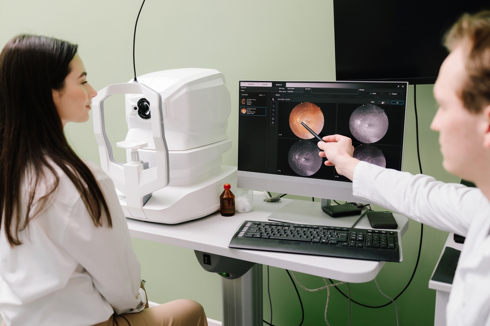
What Happens During a Retinal Scan
The process is much less dramatic than it sounds. You will sit in front of a specialized camera or scanning machine. A chin rest and forehead bar help you stay still while the device takes high‑resolution images of the back of your eye. Some exams use wide‑field cameras or advanced scanners, which can create detailed cross‑sectional maps of retinal layers.
Sometimes, your pupils may be dilated (with eye drops) so the camera gets a clearer view. The scans themselves are painless and quick, usually just a few minutes. No part of the device touches your eye.
What the Optometrist Is Looking for
Here are some of the key signs your optometrist watches for in those images:
- Retinal blood vessel changes: Narrowing, bulging, or abnormal patterns can point to diabetes, high blood pressure, or early vascular disease.
- Optic nerve health: Swelling or thinning of the nerve can suggest glaucoma or elevated eye pressure.
- Macula issues: The macula gives you central, sharp vision. Yellowish deposits (drusen), fluid buildup, or abnormal blood vessels may signal age-related macular degeneration (AMD).
- Retinal tears or detachment: A scan may catch small tears or early detachments before you even experience symptoms.
- Structural changes over time: By comparing scans year to year, your optometrist can note subtle shifts that might indicate early disease, even if you feel completely fine.
Why This Matters More Than You Think
The retina offers a real‑time view of your eye’s inner workings. Retinal scans give your optometrist early warning signs that often show up long before you notice anything. Many serious conditions start behind the scenes.
Because the retina’s blood vessels mirror your body’s circulation elsewhere, changes can hint at cardiovascular or other systemic problems. For example, high blood pressure or stroke risk can sometimes be flagged via retinal imaging.
What You Should Ask and Do
Here’s how you can make the most of your retinal scan:
- Ask your optometrist which scan is being used (OCT, wide‑field camera, fundus photo) and why.
- Request to see your images. Some clinics show you the scan and point out what they are looking for.
- Keep your scans on file and compare them over time.
- Share your complete health history so your optometrist interprets the scans in your broader health context.
- Take action when signs are found.
A retinal scan is your optometrist’s way of looking inside your health.
For more on what your optometrist sees in a retinal scan, visit Arora Vision Associates. Our office is in Cherry Hill, New Jersey. Call (856) 406-7445 to schedule an appointment today.


MAP
© 2026 Arora Vision Associates. All Rights Reserved. Accessibility Statement - Privacy Policy - Sitemap
Powered by: 